CSU-X1 and CSU-W1
Spinning disk confocal imagers from Yokogawa Electric Corporation
The Confocal Scanner Unit is an industry standard for life science imaging. BioVision offers many unique modifications on new turnkey systems or upgrades for exisiting labs.
Upgrades to the CSU10, the CSU-21, the CSU-22, the CSU-X1, or the CSU-W1 Spinning Disk Confocal Imager
BioVision Technologies offers significant upgrades to an existing CSU-series confocal imager. Suitably upgraded and incorporating a Nipkow spinning disk, the CSU acquires confocal images at video rates, minimizing photobleaching and phototoxicity – ideal characteristics for live cell work.
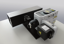
VS-Homogenizer for CSU-X1 or CSU‐W1
The Visitron Systems GmbH “VS-Homogenizer” optics are designed to enhance the laser illumination of the CSU-X1 or CSU‐W1. This optical component can be easily added to already installed confocal scan heads. The existing functionality of the original CSU confocal head remains. This enhancement offers even illumination of large sample areas and allows high‐sensitivity imaging of the specimen without the need for mathematical shading correction.
Optional Laser Merge Module
For applications requiring multiple excitation wavelengths, BioVision Technologies combines the CSU with laser merge modules from leading manufacturers. Lasers are available in a range of wavelengths and powers.
Optional Emission Filter Wheel
BioVision Technologies offers and supports filter wheels from Applied Scientific Instrumentation, Ludl Electronics Products, Prior Scientific and Sutter Instrument Company.
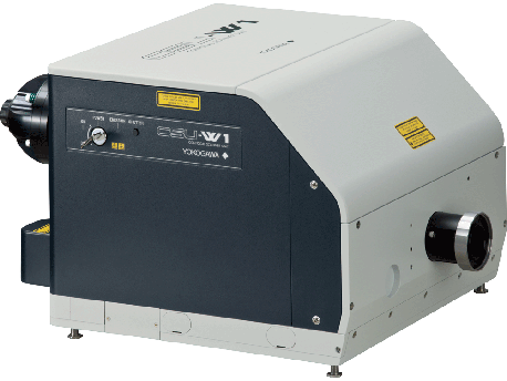
CSU-W1 Spinning Disk Confocal Imager
BioVision Technologies offers the CSU‐W1 confocal imager. Incorporating a Nipkow spinning disk, it brings a wider field of view and clearer images to the Yokogawa Electric CSU line.
With its significantly larger field of view, decreased crosstalk, and extended near-infrared spectral range, it can obtain sharper images of regions deeper inside live cells.
Features of the CSU‐W1
- Wide field of view: four times that of the conventional model of CSU (CSU10, CSU‐21, CSU‐22, CSU‐X1)
- Clearer images: a newly‐designed disk unit offers significantly reduced pinhole crosstalk, enabling clear observation much deeper into thick samples.
- Near Infrared (NIR) Port : Up to 785 nm
- Available in one‐camera, two‐camera and split‐view models
- Brightfield imaging provided with all CSU‐W1 units, via motorized mechanism for moving disks out of light path
- Pinhole size options: 25 µm pinhole disk, 50 µm pinhole disk, or dual disk with motorized disk exchange mechanism
- External light path
- 10-position filter wheel(one‐camera model, two‐camera model)
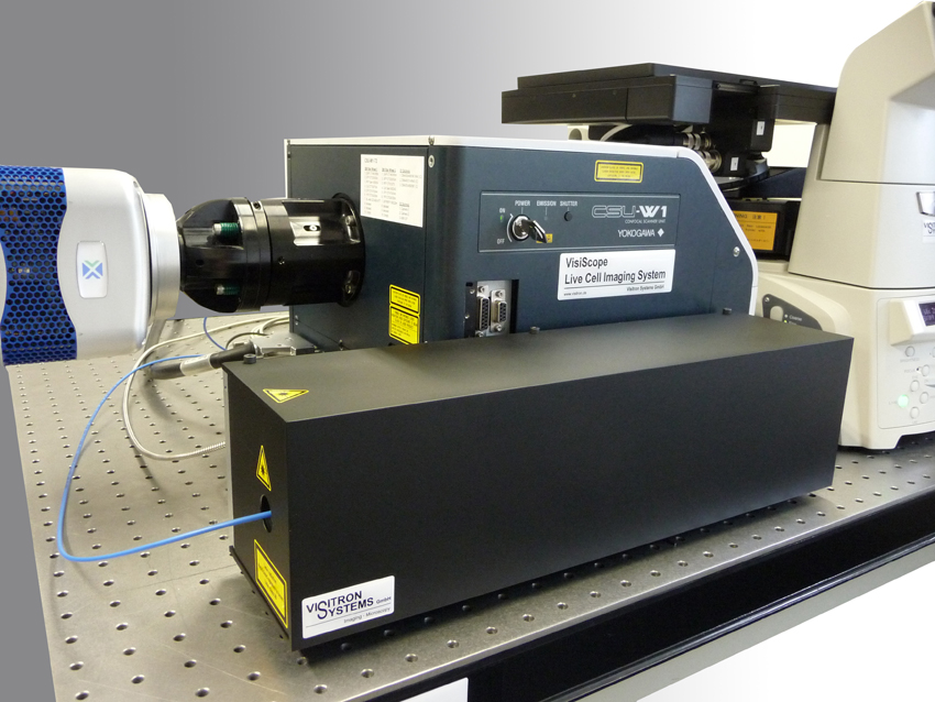
VS-Homogenizer for the CSU-W1 Spinning Disk Confocal Imager
BioVision Technologies offers the VS-Homogenizer optical component from Visitron Systems GmbH. It is designed to enhance the laser illumination of the CSU-W1.
This enhancement offers even illumination of large cell areas and allows high-sensitivity imaging of living cells without the need for mathematical shading correction.
This optical component can be added on request to CSU-W1 confocal scan heads that have already been installed.
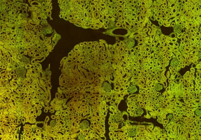
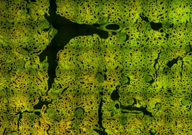
Sample Acquisitions
The following images were made by stitching images of the 13x13 mm field of view of a sCMOS camera and 63×/1.4 N.A. Oil Objective
CSU-X1 Spinning Disk Confocal Imager
BioVision Technologies offers the CSU‐X1 confocal imager. Incorporating a Nipkow spinning disk, it acquires confocal images with minimal photobleaching and phototoxicity, making it ideal for live cell work.
Basic and High‐end models are available. The Basic model offers a fixed‐speed scanner motor, while the High‐end model offers a variable speed motor.
The imaging speed of the Basic model matches that of the CSU-10, while the High-end model offers almost seven times that speed.
Its filter wheel switches between adjacent positions in 33 ms.
Features
| Type | High‐end Model | Basic Model | ||
| Spinning Speed | Standard: | 1,800 RPM (360 frames per second) | ||
| Standard: | up to 5,000 RPM (1,000 frames per second) | Optional: | 1,500 up to 5,000 RPM (1,000 frames per second) | |
| Optional: | 1,500 up to 10,000 RPM (2,000 frames per second) | Optional: | 1,500 up to 10,000 RPM (2,000 frames per second) | |
| External Synchronization | Scan‐speed synchronization through TTL pulses at rate of 300 up to 2 kHz (spinning rate of 1,500 up to 10,000 RPM) | Requires Control Unit for control of disk rotation speed | ||
| Excitation Wavelength | 450 up to 647 nm | 450 up to 647 nm | ||
| Dichroic Mirror | Optional | Automated three-channel | Optional | Manual single-channel |
| Emission Filter Wheel | Optional | Twelve-position | N/A | |
| Six-position | ||||
| External Control | RS-232C Serial Interface | N/A | ||
| Microscope Mount | C-Mount Adapter | C-Mount Adapter | ||
VS-Homogenizer for the CSU-X1 Spinning Disk Confocal Imager
BioVision Technologies offers the VS-Homogenizer optical component from Visitron Systems GmbH. It is designed to enhance the laser illumination of the CSU-X1.
This enhancement offers even illumination of large sample areas and allows high-sensitivity imaging of living cells without the need for mathematical shading correction.
This optical component can be added on request to CSU-X1 confocal scan heads that have already been installed. The existing functionality of the original CSU confocal head remains.