Light Sheet Microscopy
Improved fluorescence imaging using planar illumination
Light sheet systems are a novel method for imaging live samples. In light sheet microscopy, a sample is illuminated perpendicularly, rather than parallel to the imaging axis (as with traditional widefield or confocal microscopy).
BioVision is working with multiple manufacturers to design cutting edge light sheet systems.
Mizar TILT
The Mizar TILT is a light sheet illumination system for fluorescence microscopes. It provides all the advantages of light sheet imaging with minimal photodamage, bleaching, or toxicity. It allows imaging of epifluorescence with high‐magnification and high‐numerical aperture objectives for maximum resolution, and supports all imaging modalities including widefield fluorescence and DIC.
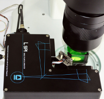
L‑SPI
The L‑shaped Single Plane Illuminator (L‐SPI) is a flexible, low‐cost illumination module enabling ultra-low phototoxicity imaging of large specimens.
This simple and effective instrument recombines two uniform, wide light sheets at right angles, reducing shadowing without the need for sample rotation or image fusion.
The system offers optional sample chambers, a fast long‐travel piezo stage and a second L‑SPI head to bring sheets in from four directions. Its compact size and large freedom of access (e.g. for positioning micro‑electrodes) make it a highly flexible solution. These strengths allow adaptation of the L‑SPI for a wide range of experimental purposes.
It is designed to work with any macro‐ or microscope, single‑mode fibre laser source and scientific camera.
The L‑SPI was co-developed by Dr. Philippe Laissue of the University of Essex and Cairn Research, Ltd..
Key benefits
- Live‐cell imaging with ultra‐low phototoxicity
- Spatial and temporal fractionation of illumination
- Orthogonal light sheets reduce shadowing
- Uniform light sheets for macroscopic observation of large samples down to individual cells
- Precise, fast and long‐travel piezo stage
- Can use your existing microscope, camera, and laser source
- Single‐ or multi‐mode FC/PC or SMA adapter
- Electro‐opto‐mechanical solution — no complex software processing
Applications
- Time‐lapse imaging with minimal phototoxicity
- Morphology of large samples (up to 10cm3)
- Confocal imaging
- Developmental biology
- Coral
- Adult zebrafish
Specifications and Options
- Single or dual L-SPI heads generate two or four orthogonal sheets
- 22um‐ or 11um‐thick sheets as standard
- 1.6mm confocal range with 22um sheet
- X-Y field of view 16x16mm
- Continuous sweeping of focal plane for reduced power density and improved homogeneity
- Fast precise piezo stage
- 20mm of Z‐axis travel, 10 mm/sec speed, 10 nm minimum step size
- Magnetic mounting platform for single or dual L‐SPI
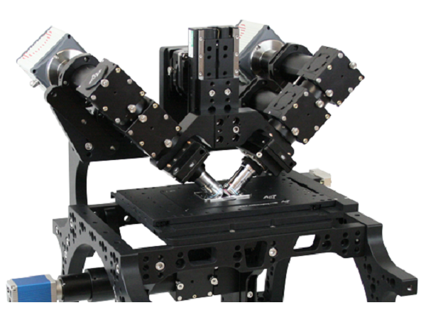
diSPIM
The diSPIM is a flexible and easy-to-use implementation of Selective Plane Illumination Microscopy (SPIM) that allows for dual views of the sample while mounted on an inverted microscope. The diSPIM was co-developed by the NIH-based lab of Hari Shroff and Applied Scientific Instrumentation (ASI).
As an ASI distributor, BioVision has hands-on experience building diSPIM systems. Please contact us with any questions about this innovative technology.
OpenSPIM
OpenSPIM is an open source platform developed to let scientists build their own low cost light sheet systems.
If you are interested in building your own OpenSPIM system, BioVision can help source all necessary components.