Application of 3D Cell Explorer to Cytology and Cytopathology
Analysis of a Drop of Blood
On this page you can explore the analysis of a drop of blood as we see it: in 3‐D and stain‐free.
The 3D Cell Explorer allows for:
- Counting white blood cells;
- Checking motility of white blood cells;
- Red Blood Cells (“RBC’s”) morphological features control.
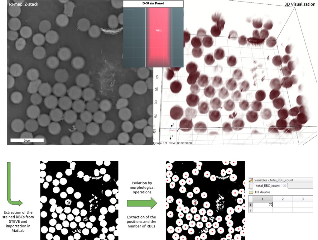
Refractive Index Map of Z‐stack, 3‐D Visualization of Blood Sample, Workflow for Analysis via MatLab
The collection of vials of blood via long, sharp needles is no longer necessary for a health check‐up. New clinical tests require only a single drop of blood! All around the world, researchers have developed less invasive, cheaper and more personalized methods of blood testing which rely on a pin prick and a small amount of blood from your finger. Using these new methodologies, up to 25 tests can be run at the same time, looking for markers related to cardiovascular diseases, diabetes and also cancer!
Nanolive wants to be part of this revolution: the 3D Cell Explorer allows an ultra‐fast, three‐dimensional and marker‐free view of the living cells inside a blood sample.
Analysis of a Drop of Blood: Counting Red Blood Cells (“RBC’s”)
A sample of human blood was taken from a donor, smeared on a coverslip and imaged at room temperature. The images were then analyzed via MatLab to determine cell count and positions.
Microbial Infections
On this page you can explore microbial infections as we see them: in 3‐D and stain‐free.
The 3D Cell Explorer allows for:
- Detection of malaria parasites;
- Study of the Life Cycle of the malaria parasite;
- Label-free detection of Chlamydia infection.
Despite the extensive use of antibiotic and vaccination programmes, infectious diseases (induced by bacteria, viruses, fungi, or parasites) continue to be a leading cause of morbidity and mortality worldwide. One of the principal tools in the diagnosis of infectious disease is microscopy. Virtually all of the isolation techniques require a microscopic examination at some point for definitive identification of the infectious agent.
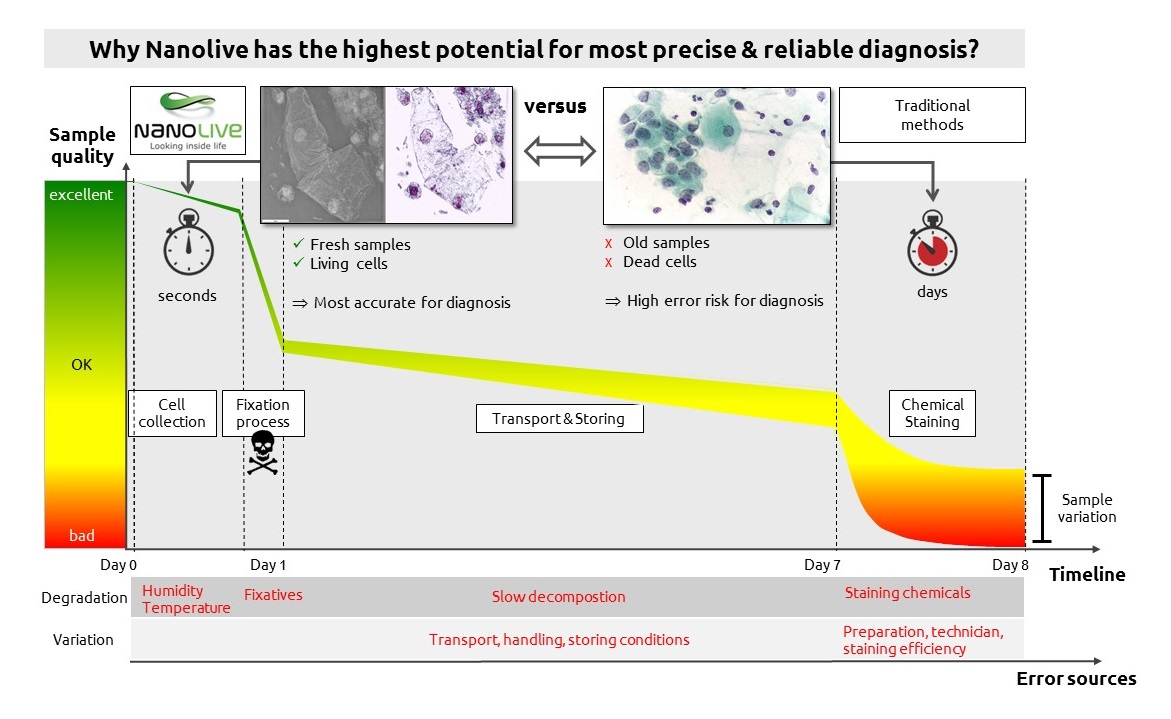
The usual routine consists of staining the specimen (with specific chemical markers) in order to give the parasites a distinctive aspect. However, there are some drawbacks: the staining and interpretation processes are labor‐intensive, time‐consuming, and require considerable expertise and trained workers.
These limitations could be solved thanks to the 3D Cell Explorer. Using only untreated samples — ones that are not fixed and not stained — Nanolive’s technology is able to identify in a few seconds the cells that have been infected by parasites, allowing their discrimination from uninfected cells.
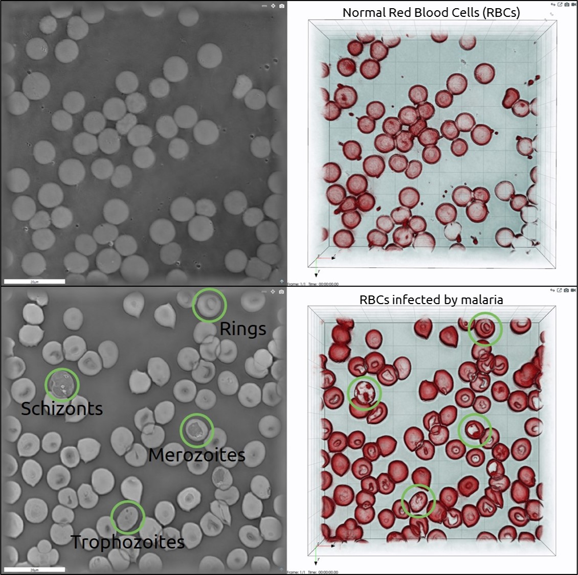
Malaria detection in Red Blood Cells (“RBC’s”)
A sample of blood infected with Plasmodium malariae was fixed and imaged using the 3D Cell Explorer.
HeLa cells infected by Chlamydia parasites
Living HeLa cells infected by Chlamydia trachomatis were imaged with the 3D Cell Explorer. The parasite inclusion bodies are displayed in green inside the cell cytoplasm.
Fast Analysis of the Pap Smear
The 3D Cell Explorer breaks every record: without any staining, the waiting time for Pap test results can be reduced from two weeks to three seconds!
Exfoliative cytopathology is an easily‐performed, noninvasive, and inexpensive procedure used in screening for preinvasive and invasive cancer cells. The incidence of cervical cancer has decreased by more than 50% in the past 30 years. That is mainly due to the increased use of a cytopathology screen called the Papanicolaou test, better known as a Pap test. It involves exfoliating cells from the cervix and smearing them on a glass slide. In order to examine them under a common microscope, the smeared biological sample must be fixed and stained. If not performed well, these procedures will compromise the test results. Thanks to Nanolive’s technology, the Pap test screening can be done in real‐time with no need for fixation and chemical staining. Preserve the natural status of cells to obtain the most precise and reliable results possible!
Marker-free 3‐D Pap Test
A fixed Pap smear slide was analyzed using the 3D Cell Explorer. The images were acquired the day after the sample collection at room temperature and without any chemical marker. Analysis conducted on a duplicate of this sample using the routine methodology confirmed our diagnosis hypothesis of mild inflammation.
Cancer Diagnosis
The 3D Cell Explorer allows for real‐time and non‐invasive cell division monitoring. The microscope detects the refractive indices (“RI’s”) in 3‐D of each and every cell organelle at a precision higher than 0.001 and with unprecedented nanometer resolution (Resolution in X and Y is 200 nm; in Z, it is 400 nm).
More and more scientific publications are pointing out how the cellular and tissue Refractive Index (“RI”) distribution is a valuable intrinsic marker for a new generation of label‐free cancer detection, more accurate diagnosis, and high‐sensitivity screening. Nanolive’s 3D Cell Explorer microscope, allowing a quantitative RI measure in 3‐D and real‐time, has the potential to perform a totally new, marker‐free and ultra‐rapid cancer detection.
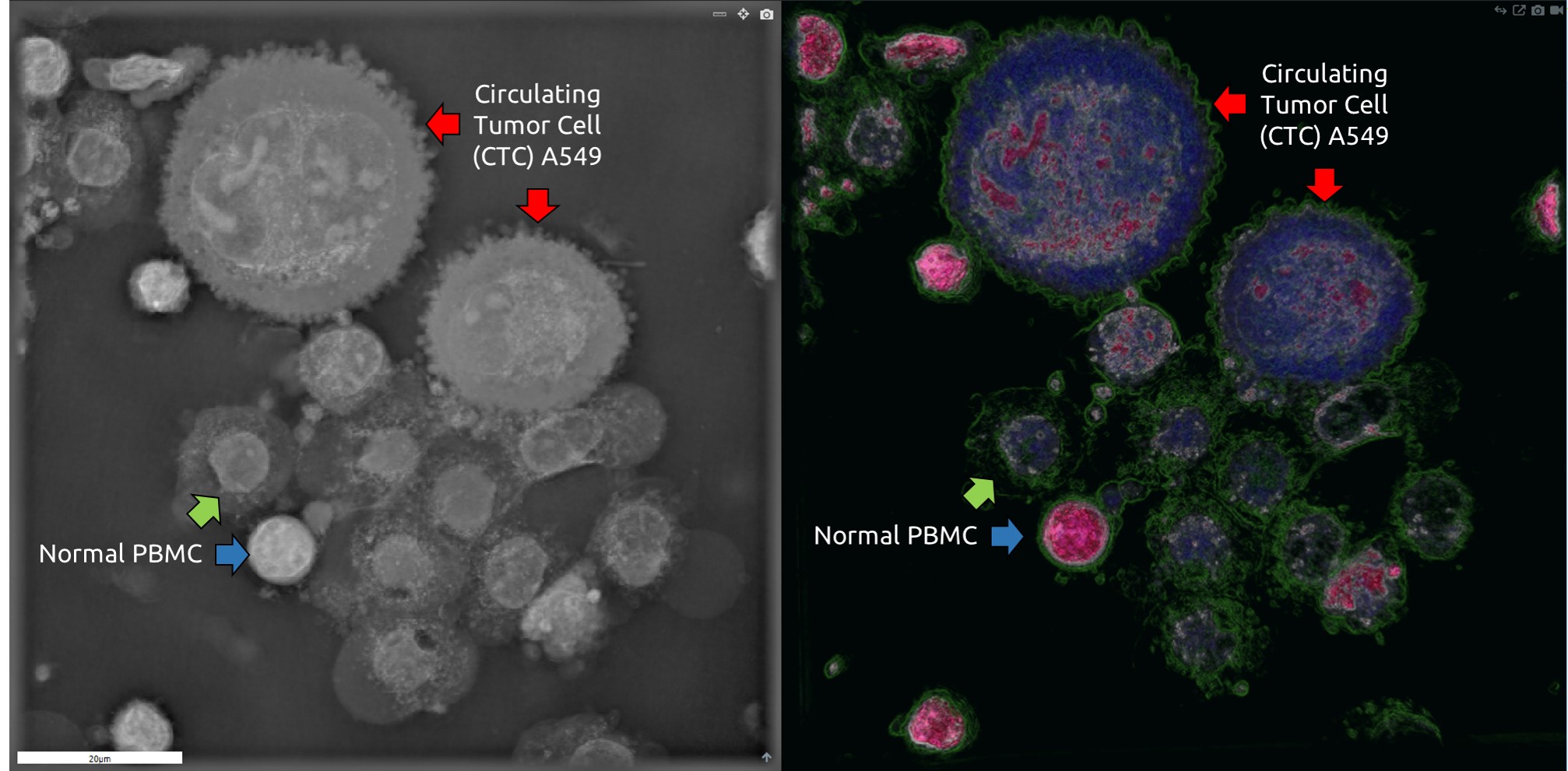
Detection of A549 Circulating Tumor Cells
Whole blood sample spiked with A549 lung cancer cells and processed on the Parsortix instrument (ANGLE plc) in order to enrich for Circulating Tumor Cells (“CTC’s”). The output of the Parsortix instrument (Peripheral Blood Mononucleated Cells, or “PBMC’s”, and A549) was resuspended in PBS and imaged by the 3D Cell Explorer.