Sepharose Hydrogel Bead - Excerpt from Z‐Series
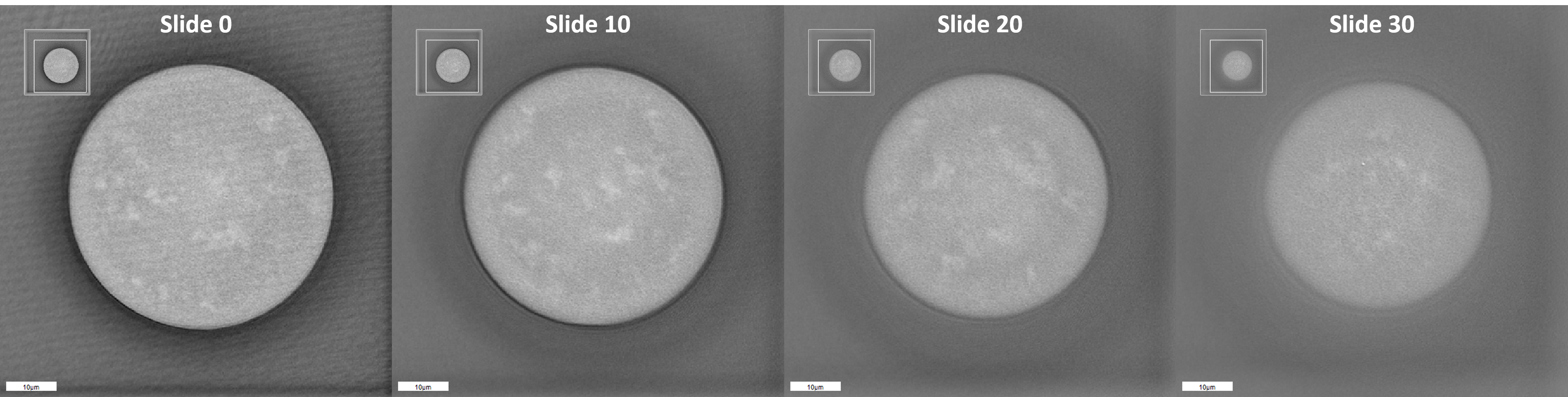
Four slices from a 96‐layer tomography acquisition of a hydrogel sepharose bead obtained by the 3D Cell Explorer. 12‐micron distance along focal axis between the first slice and the fourth.
Structural imperfections appear as the brighter spots inside the gray structure.