Application of 3D Cell Explorer to Interactions of Cells and Inorganic Materials
Hydrogels
Hydrogel products constitute a group of polymeric materials, the hydrophilic structure of which renders them capable of holding large amounts of water in their three-dimensional networks. Hydrogels are becoming more and more popular as platforms for three‐dimensional (3‐D) cell culturing. 3‐D hydrogel matrices have been used for a variety of applications, including tissue engineering of micro‐organ systems, drug delivery, cytotoxicity testing, and drug screening.
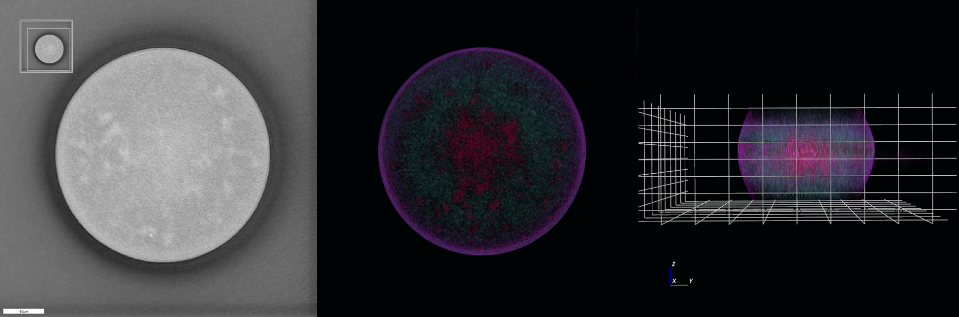
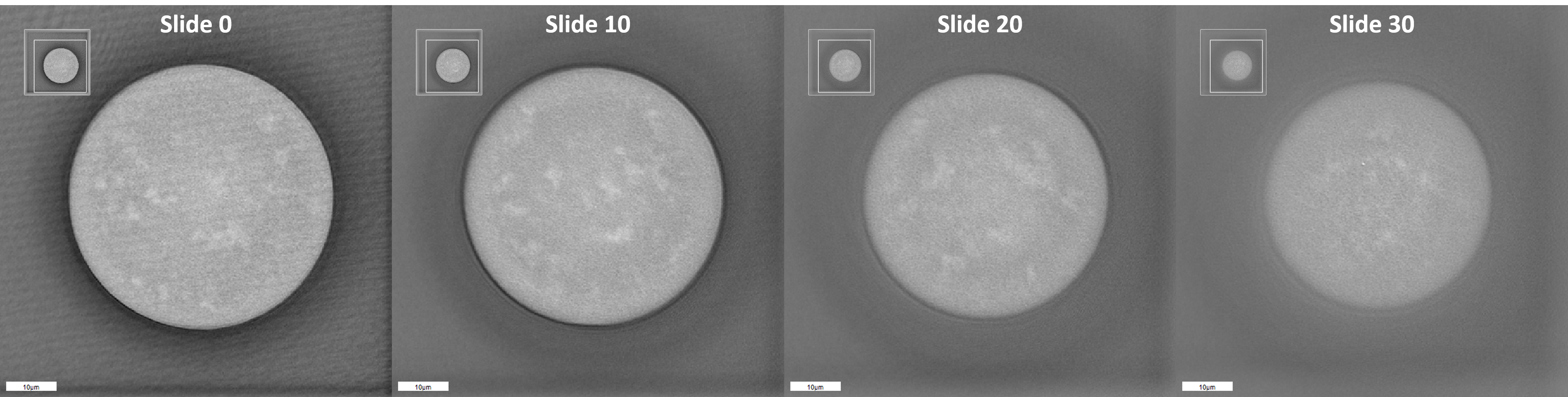
Although we imagine gel as a perfect network, in reality, due to the polymeric domains created by crosslinking, in the gel microstructure, hydrogels are not homogenous. Voids in the microstructure of the gel where a crosslinking agent or monomer has aggregated during polymerization can cause a solvent to diffuse into or out of the hydrogel. The microstructure of hydrogel therefore is not constant, and imperfections occur where water from outside of the gel can accumulate in these voids.
Although we imagine gel as a perfect network, in reality, due to the polymeric domains created by crosslinking, in the gel microstructure, hydrogels are not homogenous. Voids in the microstructure of the gel where a crosslinking agent or monomer has aggregated during polymerization can cause a solvent to diffuse into or out of the hydrogel. The microstructure of hydrogel therefore is not constant, and imperfections occur where water from outside of the gel can accumulate in these voids.
Nanolive’s technology provides a new approach to check in real‐time the morphological features of these hydrogels: the 3D Cell Explorer allows one to obtain a complete tomography of these structures pointing out all the imperfections and localize them in 3‐D.
Membranes
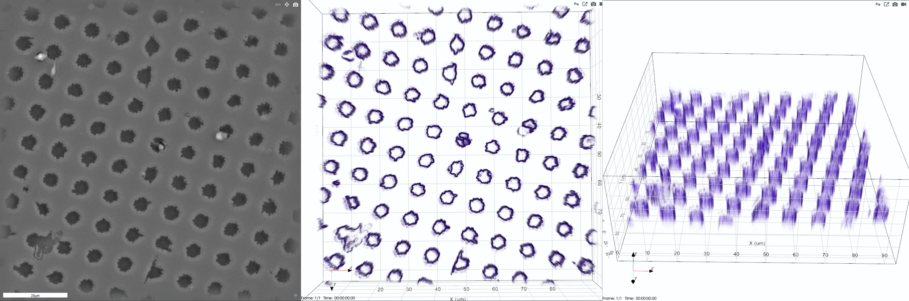
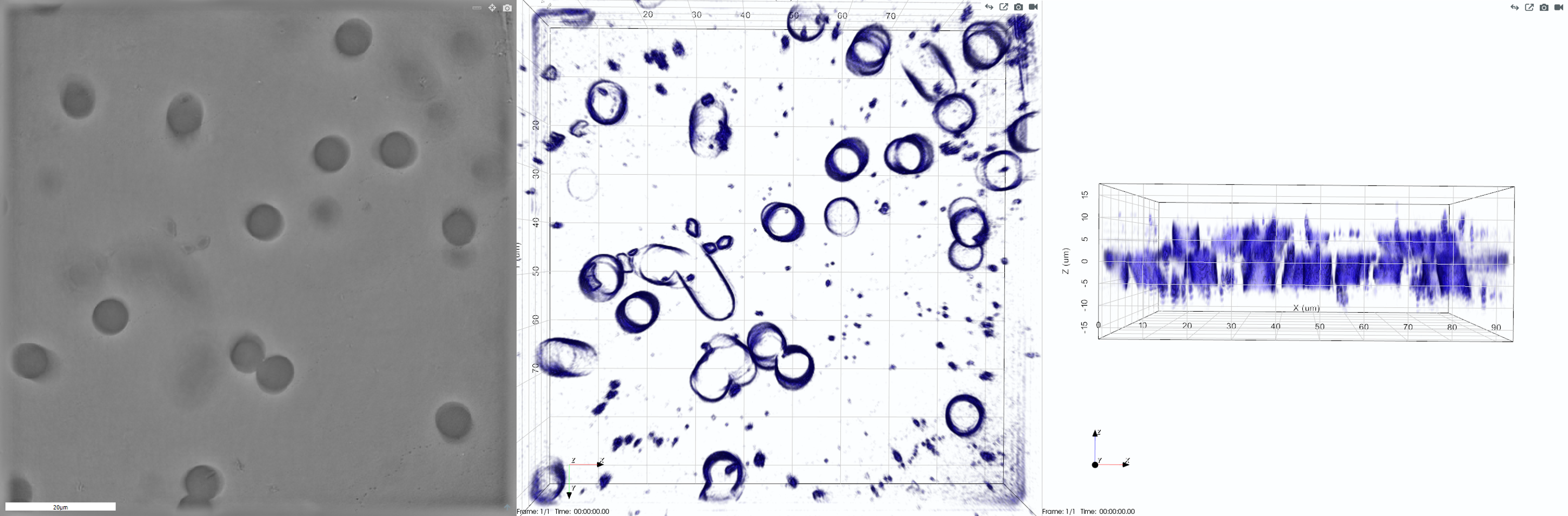
Porous membranes are intended for separation of larger molecules such as solid colloidal particles, large biomolecules (proteins, DNA, RNA) and cells from the filtering media. They are used in microfiltration, ultrafiltration, and dialysis applications.
Usually we imagine membrane pores as parallel, nonintersecting cylindrical capillaries. In reality the structure of pores can be a random network of unevenly‑shaped structures of different sizes.
The 3D Cell Explorer can be used to obtain crucial morphological information about their structure, thanks to marker‑free, real‑time 3‑D reconstruction.